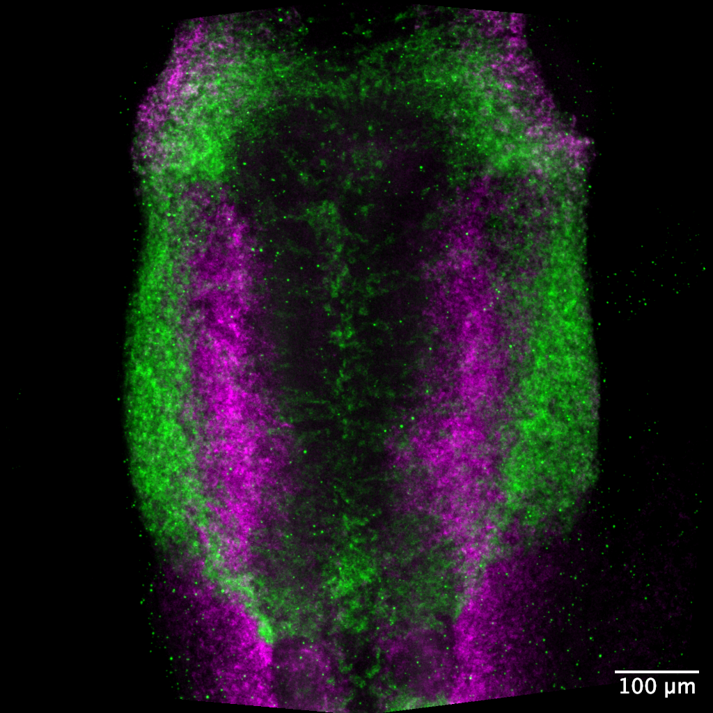
Avian neural crest migrating
Multiplexed HCR staining for Sox10 and Twist1 at HH10. Sox10 is expressed by migrating neural crest cells and Twist1 is expressed by the underlying cranial mesoderm.
Somite progenitor explant migrating
Cells are mosaically marked by electroporation of a membrane GFP construct.

Duplicated digit and feather patterns
Wing resulting from ZPA transplant to the anterior margin of the limb bud. Both the digit pattern and the feather pattern are perfectly duplicated in the AP axis.
C. elegans early cell divisions
Histones are labelled in magenta and microtubules are labelled in cyan.
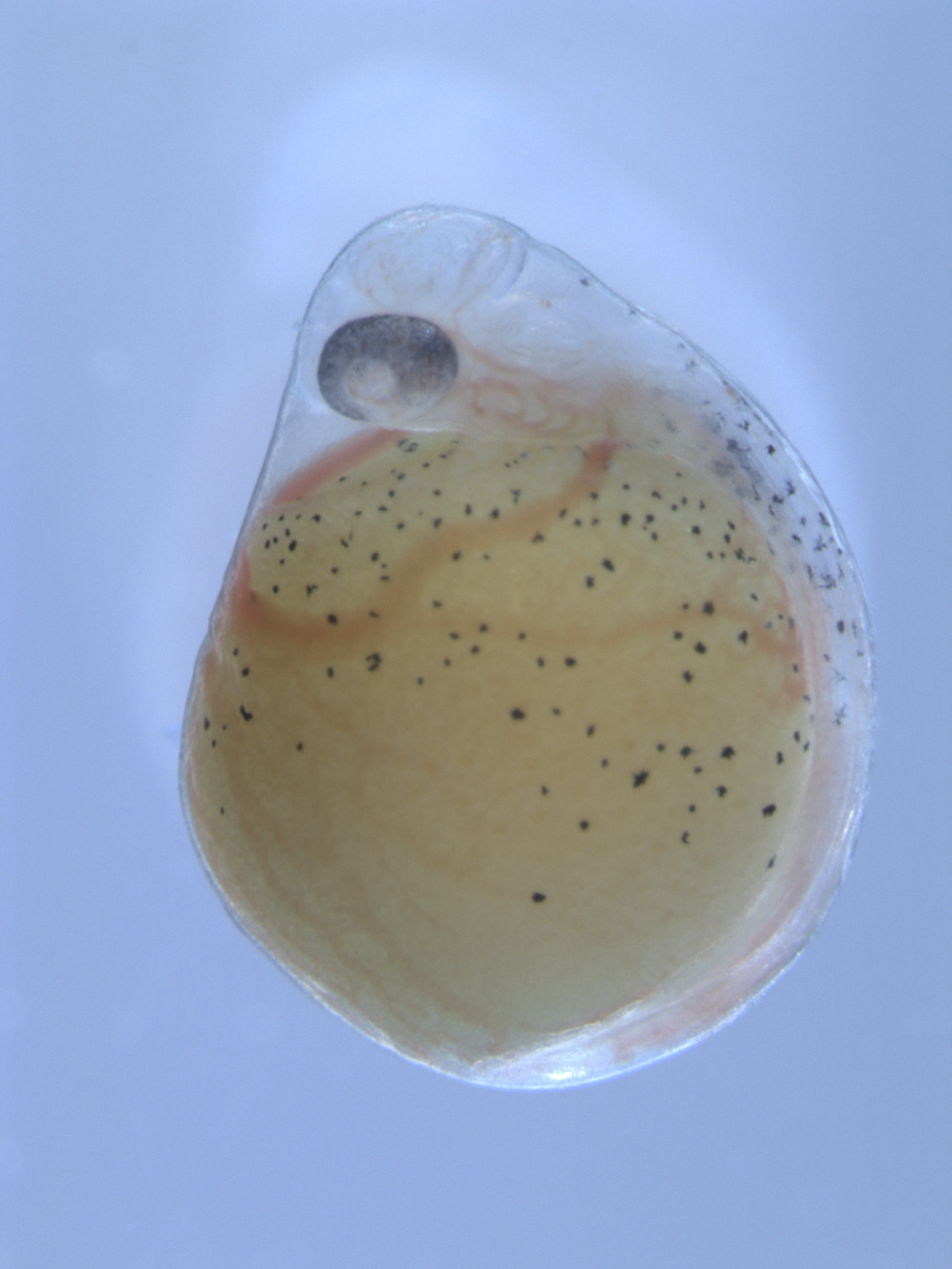
Astatotilapia calliptera embryo
Brightfield image at 7dpf. The embryo is wrapped around the large cylindrical yolk.
Xenopus laevis neural crest explant
Neural crest cells from membrane-GFP transgenic migrating on fibronectin.
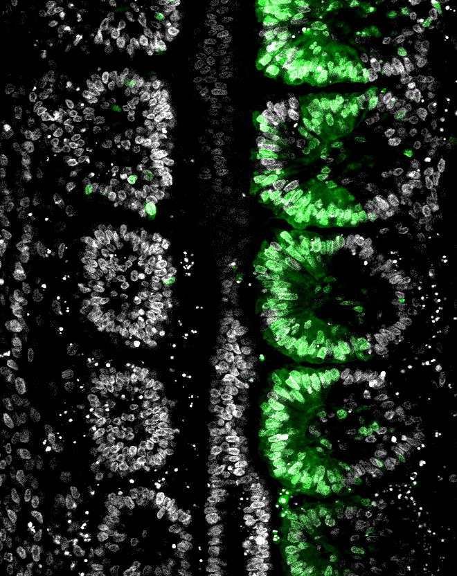
Avian somite progenitor transplant
Somite progenitor cells were transplanted from a GFP transgenic embryo into a wildtype embryo. The image shows nuclei in white and transplanted tissue in green - cells have incorporated into the medial somite.
Crepidula fornicata epiboly
Embryo injected with membrane GFP (cyan) and H2B-RFP (magenta) constructs and imaged for 24 hours.
_triple_composite_w_DAPI.jpg)
Danio rerio cranial region
24hpf embryo stained for sox10 (green), ets1 (yellow) and twist1a (magenta) transcripts.